
Destinations & habitats
Crown-of-Thorns Scar or Bleaching? Reading a Great Barrier Reef Patch
A fresh crown-of-thorns feeding scar and a bleached coral can both look white, but the edge, remaining tissue and wider reef pattern tell different stories.
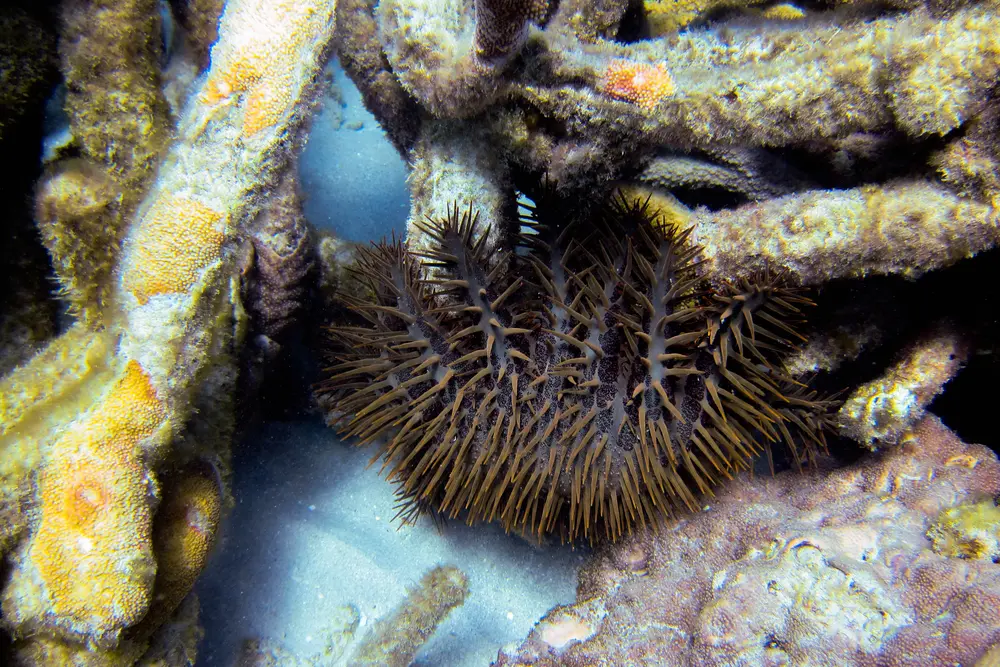
A crown-of-thorns starfish photographed at Lizard Island on the Great Barrier Reef. The frame establishes the animal and place; it does not show feeding, a fresh scar or an outbreak.
Photo: Ryan McMinds. Image source · CC BY 2.0. Full frame auto-oriented and resized from 3648 by 2432 to 1000 by 667 pixels, metadata removed and converted from JPEG to WebP; no crop.
.jpg)
On a Great Barrier Reef dive, look at the boundary of a white coral patch and ask whether living tissue still covers it: that small check matters because bleaching and fresh tissue loss can expose the same bright skeleton to your eye. A crown-of-thorns feeding scar is bare skeleton where tissue has been removed, while a bleached coral still has stressed, translucent tissue over its skeleton—even though telling the two apart from a passing view can be difficult. 1 2
Start with the patch, not a dramatic diagnosis. A sharp white area beside normally coloured tissue, especially with a crown-of-thorns starfish on or beneath the colony, is strong context for recent feeding. Without the animal, close tissue detail or repeated observations, disease, coral-eating snails and other injuries can leave similar white skeleton. Record the edge and its surroundings, but leave the cause open when the clues do not line up. 3
Start at the margin, not the colour
White is the first clue and the least decisive one. In bleaching, the coral animal loses many of the microscopic algae that normally colour and feed it. Its thin tissue becomes transparent, so the white limestone skeleton shows through. The colony is stressed but not necessarily dead. Through a clear, close view you may still see intact polyps or a continuous tissue surface, and nearby colonies may be pale or fluorescent too. Bleaching can affect a whole colony, one sunlit surface or irregular patches, so shape alone does not settle the question. 2 3
A fresh feeding scar is tissue loss. The exposed skeleton can look stark white beside brown, green or purple living tissue, often with a relatively distinct boundary. If the crown-of-thorns is still draped across the colony, the animal-plus-edge combination is much more informative than the colour alone. Check under ledges and between branches only with your eyes or camera; never reach in or turn coral to complete the view. The starfish's spines are venomous, and touching the colony would add damage to the scene you are trying to understand. 1
Even a clean boundary is not a signature. AIMS survey divers record crown-of-thorns scars, Drupella snail scars, white-syndrome disease and unknown scars as separate observations because several agents remove tissue and expose skeleton. Bleaching is recorded separately as a percentage of living coral cover. That professional distinction is a useful field habit: note whether you see colour change, tissue loss, a predator or only an unexplained white patch rather than forcing every white mark into one box. 4
Distance and water movement can hide the deciding texture. If your view is oblique, surge is moving you or the patch is high above you, keep the observation at the level you can support: pale colony, white patch, exposed-looking skeleton or starfish present. A wide photograph that includes the entire colony and its neighbours often preserves more evidence than a tight white close-up, because it shows whether the change is isolated or repeated across the reef.
The starfish leaves a meal-shaped gap
A crown-of-thorns does not scrape coral with its spines. Like other sea stars, it pushes its stomach out through its mouth and spreads that tissue across the coral surface. Digestive enzymes break down the living coral tissue, and the starfish absorbs the meal before moving on. What remains visible to a diver is the negative of that contact: a white area where tissue has gone, sometimes tracing the portion the animal could cover. 1
The starfish may be easier to miss than the result. Its many arms and dense spines break up its outline among branching coral, and an animal can shelter under a plate or in reef structure between feeding bouts. Do not interpret an empty scar as proof that the animal has left the reef. In the 2025–26 AIMS survey, observers found crown-of-thorns starfish on 27 surveyed reefs but recorded feeding scars on 77; the scars indicated that low numbers could be present even when the survey did not see the animals themselves. 5
That same result sets a limit. One scar shows that tissue was lost at one colony; it does not reveal how many starfish are present or whether the reef is in outbreak. AIMS reported no severe outbreaks in its 2026 surveys, while established outbreaks occurred on five reefs and an emerging fifth wave was evident in the north. Conditions therefore differ among reefs and through time. Use the current local briefing and formal monitoring, not a single photograph, for reef-scale status. 5
Preferred prey can also bias what catches your eye. At Lizard Island, a 2023–2025 field study found Acropora made up more than 80 percent of recorded crown-of-thorns diet even as coral cover and individual feeding rates changed after mass bleaching. On a dive, a cluster of white Acropora branches may therefore mix two disturbances in one view. Record surrounding bleaching and visible starfish separately; do not subtract one cause from the other by appearance alone. 6
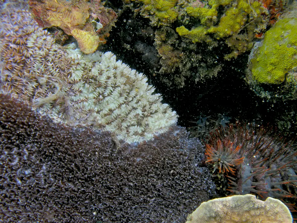
In this documented Timor-Leste feeding scene, a crown-of-thorns starfish lies beside a sharp boundary between living coral tissue and exposed white skeleton. The image is not from the Great Barrier Reef, and it does not make every pale patch a feeding scar.
Photo: Nick Hobgood. Image source · CC BY-SA 3.0. Full frame auto-oriented and resized from 2695 by 2021 to 1000 by 750 pixels, metadata removed and converted from JPEG to WebP; no crop. The adapted image remains available under CC BY-SA 3.0.
_starfish_and_hard_coral_damage_after_feeding.jpg)
A white scar does not stay white
A fresh scar is a moment in a longer sequence. In a 2026 study at One Tree Reef, researchers followed 71 discrete scars made by individual crown-of-thorns starfish in a low-density population. Forty-six scars retained living coral tissue around their edges. Turf algae commonly occupied the exposed surface first, and crustose coralline algae appeared on some scars by ten weeks. All tracked scars kept their skeletal structure through the 22-week study, but their colour and cover changed. 7
Living edges mattered, but they did not guarantee repair. Of the 46 scars with tissue remnants, 26 percent showed coral growth over at least part of the scar by week 22; the other 74 percent did not. None of the 25 completely denuded colonies showed regeneration in that period. The observed regrowth was concentrated in Isopora, Montipora and Porites, while the tracked Acropora scars remained covered by turf or crustose coralline algae. Those are results from two One Tree Reef sites, not a timetable for every coral you meet. 7
For a diver, the visible translation is that age changes the clue. Bone-white skeleton beside intact tissue suggests recent loss. A duller green-brown film or pink crust suggests colonisation after exposure, but it cannot by itself name the original cause. Coral-coloured tissue creeping inward would be evidence of regeneration only if the same scar were relocated and photographed consistently. Two similar patches on different colonies are not a before-and-after pair.
Bleaching has its own sequence: living tissue may regain its algal partners and colour if stress eases, or it may die and later become overgrown. That convergence is why an old scar, a dead bleached patch and some disease lesions can all lose the bright-white appearance that first caught your eye. Notice what is present now—tissue, algae, a sharp margin, nearby pale colonies—rather than reconstructing an unobserved history from colour alone. 2 3
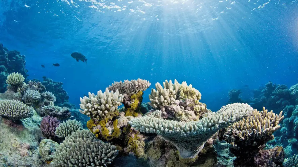
NOAA identifies this frame as healthy corals beside corals showing signs of bleaching. It demonstrates a reef-scale colour pattern, not a Great Barrier Reef site or proof of what caused any one white patch.
Photo: NOAA. Image source · Public domain — U.S. NOAA. Full frame resized from 1195 by 670 to 1000 by 561 pixels, metadata removed and converted from PNG to WebP; no crop.

Record the patch and leave the verdict open
If a white patch deserves a second look, hold position without settling onto coral. Take one wide frame that includes the colony and neighbouring reef, then a closer frame from the same safe distance if visibility allows. Include the starfish in the wide image when one is already visible; do not chase it under coral or move anything. Note the date, approximate depth, reef or site name, patch location on the colony and whether the surrounding corals also looked pale.
Photographs can preserve useful distinctions that memory drops after surfacing. A scale reference that is already in the scene—branch width, the whole colony or your camera's unchanged field of view—is better than placing an object beside the coral. White balance may improve consistency, but it cannot restore missing tissue or prove that tissue is present. Keep the original file as well as any corrected version, and describe what you observed before assigning a cause.
Visitors can submit photographs of crown-of-thorns starfish, coral bleaching and other reef sightings through the Reef Authority's Eye on the Reef app. The categories are separate, which is another reason to report the strongest visible clue rather than a confident guess. A location and wide context can help trained observers evaluate a record; one anonymous white close-up cannot establish an outbreak. 8
Use the Great Barrier Reef regional guide to place the encounter without treating the map as a sighting promise, and compare the scar's edge with the way a mushroom coral moves across sand or how distance changes underwater colour. On your next reef dive, let the first white patch slow your eyes: look for remaining tissue, the line of the edge, a hidden starfish and the pattern on nearby colonies. The best observation may end not with a diagnosis, but with a precise record of what the reef actually showed you.
Sources & further reading
- Reef Authority — Coral predator outbreaks — Great Barrier Reef Marine Park Authority institutional account. Supports the everted-stomach feeding mechanism, exposed white skeleton and venomous-spine safety point.
- AIMS — What is coral bleaching? — Australian Institute of Marine Science explainer. Supports bleaching as transparent living tissue over white skeleton, possible recovery and variable colony-scale patterns.
- NOAA CDHC — Discolored Tissue and Bleaching — NOAA Coral Disease and Health Consortium field guide. Supports the distinction between colour change with tissue present and recent tissue loss, plus the warning that bleaching and white lesions can be confused.
- AIMS — Reef monitoring sampling methods — AIMS monitoring protocol. Survey divers record crown-of-thorns, Drupella, disease and unknown scars separately from bleaching; supports the observation-first field categories.
- AIMS — Annual Summary Report of Coral Reef Condition 2025/26 — AIMS Annual Summary Report 2025/26, accessed September 28, 2026. Supports the current reef counts, the difference between animal detections and scar detections, and the qualified outbreak context.
- Levering et al. (2026), Changes in feeding behavior of Pacific crown-of-thorns starfish following mass coral bleaching — Coral Reefs, published 2026; primary 2023–2025 Lizard Island field surveys. Supports the site-specific Acropora diet share and the need to record starfish density, feeding and bleaching as distinct observations.
- Foo et al. (2026), The Fate of Coral Feeding Scars Following Predation by Individual Crown-of-Thorns Seastars — Ecology and Evolution 16, e74285; DOI 10.1002/ece3.74285; first published September 23, 2026. Open primary study of 71 discrete feeding scars at two One Tree Reef sites; supports remnant, colonisation and 22-week regeneration results without extending them to outbreak feeding.
- Reef Authority — What is Eye on the Reef? — Great Barrier Reef Marine Park Authority citizen-observation guidance. Supports photo submissions for crown-of-thorns starfish, bleaching and other reef sightings.