Ocean discoveries
How a Mushroom Coral Walks Toward Blue Light
A small mushroom coral resting on sand can shift its whole body toward blue light, turning a seemingly fixed part of the reef into an animal whose position is worth watching.
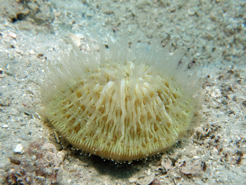
Cycloseris cyclolites photographed by Lyle Vail at 18 metres off Lizard Island, Australia, in 2014. Notice the separate disc resting on sand and the living tissue over its ridges; this field photograph identifies the subject but does not document movement.
Photo: Lyle Vail. Image source · CC BY 3.0 Unported. Unmodified original image; article retains full frame. Archive thumbnail uses a display crop.

Look for a small, round mushroom coral resting on sand, with ridges radiating toward its central mouth. That apparently stationary disc may be capable of moving its whole body: research on Cycloseris cyclolites shows how swelling and twisting tissue can carry a free-living coral toward blue light. 1 2
The answer is not a hidden set of legs. In a study published on January 22, 2025, Brett Lewis and colleagues used laboratory time-lapse imaging to reveal movement that is easy to miss during a dive. Soft tissue changes shape around a hard skeleton, makes contact with the bottom, and helps shift the animal. The discovery gives you something new to look for beside the reef, without promising that a coral will cross your view while you wait. 1
Find the disc, then separate swelling from travel
Begin with the gap around the animal. A free-living mushroom coral lies on the bottom rather than forming a branch attached to the reef framework. In the photograph above, sand surrounds a distinct circular body. Follow the ridges inward toward the mouth, and look for the translucent tentacles above them. The stone-like shape is not the whole animal: living tissue covers the skeleton.
Corals of the World describes Cycloseris cyclolites as a small circular or slightly oval dome, often cream or greenish, with straight, symmetrical skeletal ridges called septa. That term earns its place when you follow the lines toward the mouth. It does not make a round outline a reliable species identification: the account treats this coral as a species complex and warns that many western Indian Ocean and Red Sea records may represent something else. Keep uncertain sightings at 'mushroom coral' rather than assigning this name from shape alone. 2
Now compare panels A and B below. The coral becomes taller and fuller as its tissue inflates; in C through E, tissue around the edge twists during movement. By F, it has deflated again. Those changes explain why an outline can mislead you. An edge extending over a grain of sand could be swelling outward while the centre stays in place, not the entire coral traveling across the bottom. 1
If you take a photograph, include the mouth and a fixed piece of surrounding reef in the same frame. A later image from a similar angle gives you a better comparison than two close-ups with no shared background. Do not place a marker beside the coral or rearrange loose rubble to create a reference. If the mouth is hidden, record that limitation; a changing silhouette is still an observation, but it answers a different question from a changing position.
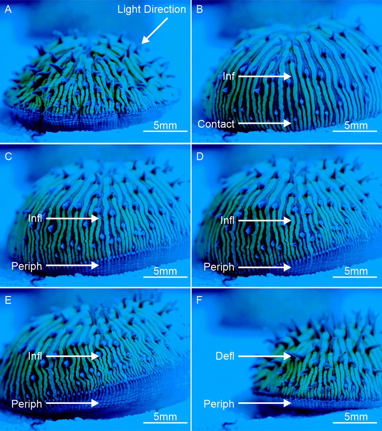
Laboratory sequence from Lewis and colleagues, Figure 3. Compare the lower profile in A and F with the expanded tissue in B–E; the arrows identify inflation and peripheral tissue involved in movement. The 5 mm scale bars show how small the subject is. These are selected research images, not six successive moments visible at normal diving speed.
Photo: Brett M. Lewis, David J. Suggett, Peter J. Prentis and Luke D. Nothdurft / PLOS ONE (2025), Figure 3. Image source · CC BY 4.0 International. Complete publisher figure, unmodified; all panels, labels and scale bars retained.
Blue light is a direction—not a starting signal for divers
The blue cast that builds up between your camera and a distant reef is part of this story. Water changes the light available underwater, and our guide to underwater colour and white balance follows what that does to a photograph. Here the question shifts from what your camera records to whether an animal on the bottom responds differently to different light.
Lewis and colleagues compared movement toward controlled blue and white light sources in aquaria, using five individual corals across repeated trials. Movement toward blue light occurred more often than movement toward white light. This directional response is called phototaxis: 'toward the light' describes a change in position, not simply a coral looking bluer under a lamp. The small laboratory sample matters when you return to the reef; it is evidence of an ability, not a forecast for every disc you find. 1
To see how that position changes, look at the underside sequence below. Researchers filmed through the glass bottom of an aquarium. Inflation widened contact around the edge, and tissue twisted against the supporting surface. This is the hidden counterpart of the swelling visible from above. You cannot obtain that underside view on a dive without disturbing the animal, so let the research images supply it. 1
Why might light direction matter beside a sandy reef slope? The authors propose that blue light could help these corals reach suitable deeper habitats. That is an interpretation of the laboratory response, not a route they followed with tagged wild corals. A blue-lit patch next to your subject is therefore a feature to include in the scene, not proof that the animal is heading there. 1
Light also supplies energy to photosynthetic algae living in the tissues of many reef corals. NOAA explains how those partners provide the coral with products of photosynthesis and contribute to its colour. When you look at living tissue spread over a skeleton, you are looking at the surface where that partnership operates—not at a bare stone that merely reflects light. This makes the lighting around a coral relevant, without telling you its preferred destination from a single photograph. 3
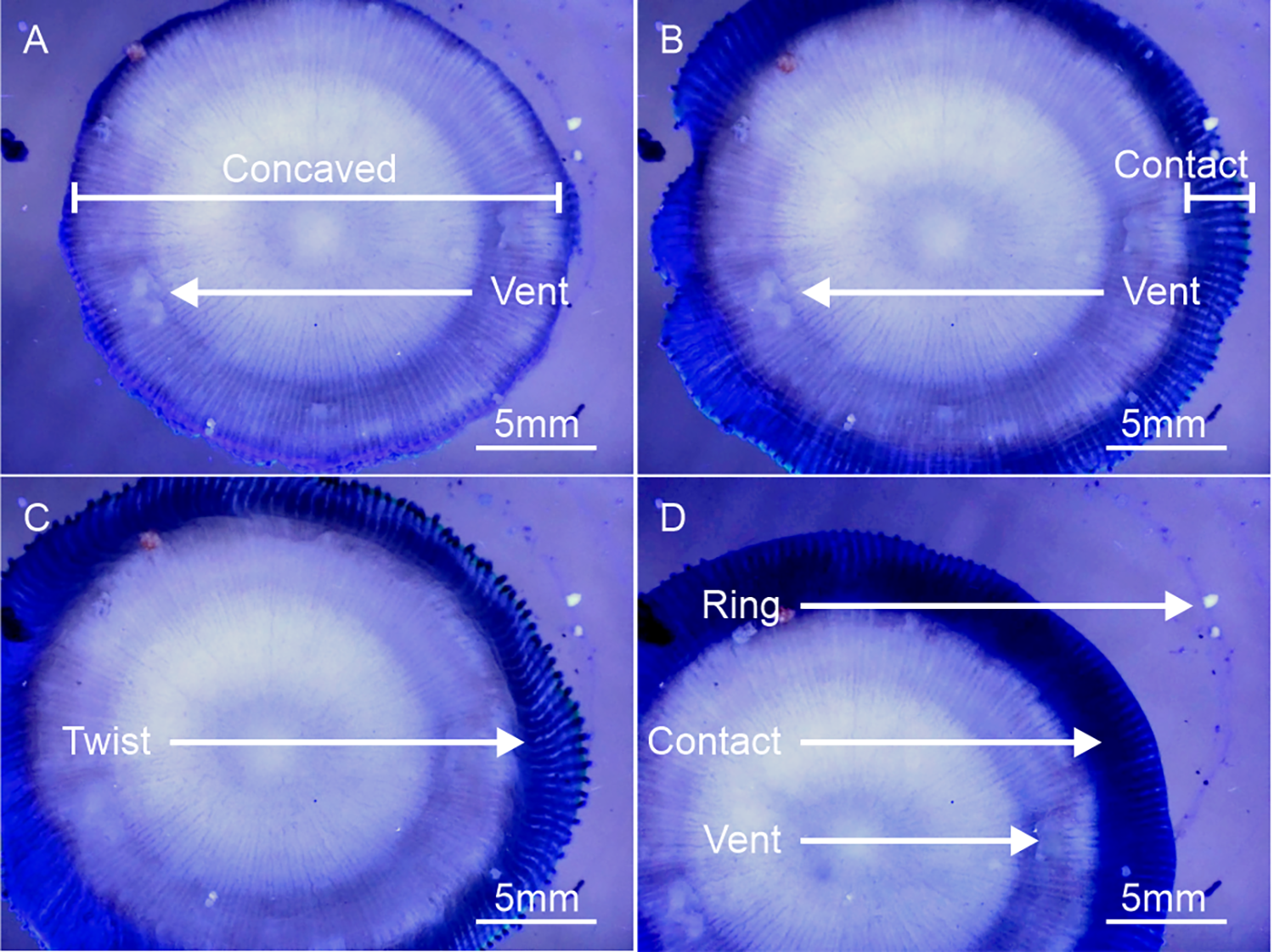
Laboratory underside views through glass, from Lewis and colleagues, Figure 4. Follow the rim from the narrow resting contact in A to the wider contact in B, then the twisting tissue in C. D shows a residual sediment-and-mucus ring. Blue light came from the lower left; this is not a view obtained by turning a wild coral over.
Photo: Brett M. Lewis, David J. Suggett, Peter J. Prentis and Luke D. Nothdurft / PLOS ONE (2025), Figure 4. Image source · CC BY 4.0 International. Complete publisher figure, unmodified; all panels, labels and scale bars retained.
A changed position does not tell you what moved it
Suppose a return photograph places a coral farther from the same rock. You have evidence of displacement, but you have not yet identified the cause. The 2025 study also recorded inflated corals being carried or rolled by water flow. In those observations, the current-driven movement did not follow the light. Active tissue movement and a passive ride can therefore produce the same broad result: a coral somewhere new. 1
A field study by David Bayley and Andrew Mogg puts that distinction on a reef rather than in a tank. They used overlapping underwater photographs to reconstruct a patch of lagoon reef in the Chagos Archipelago, then compared the positions of 51 mushroom corals between 2017 and 2018. The four species were not Cycloseris cyclolites. Most shifted downslope, but the researchers could not turn two survey snapshots into a continuous account of how each animal got there. 4
That is a useful limit for your own pair of images. A start and finish do not show whether the coral paused, changed direction, or was pushed by a surge between visits. Even identifying the same individual needs care when several similar discs share the sand. Keep a wider reference frame, note the date and depth, and compare distinctive outlines and surrounding features before calling two photographs a journey.
Swelling itself has other uses. Earlier time-lapse research documented mushroom corals freeing themselves from burial through repeated inflation. That experiment helps explain why expanding tissue beside sediment is worth noticing, but it does not establish that every swollen coral is walking or trying to escape sand. Nor is it a reason to cover one and wait for a response. Let the position, tissue shape and surrounding sediment remain separate entries in your observation until you have evidence that connects them. 5
Leave the coral where its next move can be its own
Approach a sandy gap with the same care you give the reef framework. Hover clear of the bottom and keep your fins from stirring sediment over the coral. If you cannot hold that position without settling down or gripping the reef, continue the dive. A photograph is not worth changing the conditions around the animal you want to understand.
Do not lift a mushroom coral to check whether it is attached, turn it over to find the tissue shown in the microscope images, or aim a blue light at it to prompt movement. The experimental response was established under controlled conditions. Recreating a fragment of that setup during a dive would produce a disturbance, not a useful field test.
Give the surrounding sand some space in your composition. Record what you actually see: a rounded disc, exposed ridges, extended tentacles, an inflated outline, or a change relative to a fixed landmark. If nothing changes during your visit, that is not a failed encounter. The research used time-lapse precisely because a short look can miss a slow process.
For Australian reef context, explore the Great Barrier Reef regional map; use the Cycloseris cyclolites species account to compare the identification details. The map is regional context, not a promise of finding this coral at a particular marker.
On your next dive, look past the fish moving above the sand. Find a separate coral disc, notice where its living edge meets the bottom, and keep its centre in the picture. The useful surprise is not that you can make a coral walk—it is that an animal you might have treated as scenery can change its own position.
Sources & further reading
- Lewis, Suggett, Prentis & Nothdurft (2025), Walking coral: Complex phototactic mobility in the free-living coral Cycloseris cyclolites — PLOS ONE 20(1), e0315623; published January 22, 2025. Primary laboratory study and supporting time-lapse videos. Five individuals reused across trials; habitat navigation remains an interpretation. Figures 3 and 4 reused under CC BY 4.0.
- Corals of the World — Cycloseris cyclolites — Expert species account: morphology, habitat and species-complex warning. Consulted September 15, 2026.
- NOAA — What is Zooxanthellae? — National Ocean Service explanation of photosynthetic partners in coral tissues; updated December 12, 2024.
- Bayley & Mogg (2023), Mushroom to manoeuvre? Using photogrammetry to track the movement and survival of free-living corals — Coral Reefs 42, 271–278; first published online December 13, 2022. Repeat field surveys of 51 corals from four other species in Chagos; endpoint displacement does not reveal the complete path or distinguish all movement mechanisms.
- Bongaerts, Hoeksema, Hay & Hoegh-Guldberg (2012), Mushroom corals overcome live burial through pulsed inflation — Coral Reefs 31, 399. DOI: 10.1007/s00338-011-0862-z. Primary time-lapse observation of inflation and sediment removal; not a visitor experiment to repeat.